


Foot mycosis(dermatophytosis,Tinea pedis) is a foot skin disease caused by pathogenic or opportunistic fungi.Skin changes on the legs are characterized by peeling, which is accompanied by itching.In the case of severe lesions, against the background of red and swollen skin, erosion and deep cracks appear on the soles of the feet and in the space between the toes, which are accompanied by pain and make it difficult to walk.
The code according to the International Classification of Diseases, 10th revision (ICD-10) is B35.3.
The advent of modern antifungal drugs has improved the epidemiological situation, but foot mycosis still remains one of the most significant problems in dermatovenerology.The use of some drugs is limited in the elderly and patients with chronic diseases.
Prevalence of foot mycosis.According to the World Health Organization (WHO), about 1/3 of the world's population suffers from fungal diseases, where the most common are foot mycoses;the incidence is increasing every year.
According to dermatologists, 10-20% of the adult population suffer from foot mycoses;in men this disease occurs 2 times more often than in women, and in older people more often than in young people.At the age of more than 70 years, foot mycosis is registered in every second patient, which is associated with an increase in concomitant metabolic and vascular changes (diabetes mellitus, varicose veins, etc.).Increasingly, foot mycoses are detected in children.
Millions of people are now affected by this disease.Workers in some professions are at risk: miners, athletes and military personnel.
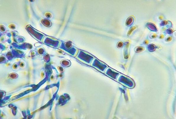
Causes of foot mycosis.The most common causes of foot mycosis are dermatomycete fungi: Trichophyton rubrum (90%), Trichophyton mentagrophytes, and less commonly Epidermophyton.Sometimes foot mycosis can be caused by fungi of the genus Candida.
Risk factors for foot mycosis:
- Exogenous (external): microtrauma of the skin of the feet (calluses, corns), cracks, increased sweating, wearing tight shoes, shoes made of artificial materials, failure to observe the rules of personal hygiene, irregular foot washing and poor drying with a towel.
- Endogenous (internal): varicose veins and vegetative-vascular dystonia, which leads to insufficient blood supply to the skin of the legs;hypovitaminosis;taking glucocorticosteroid, cytostatic, antibacterial and estrogen-progestin drugs, which reduce the body's overall immunity.
Infection with foot mycosis can occur directly from a sick person, and it can also be spread through contact and household contact (in swimming pools, baths, gyms, through shoes, towels, carpets, etc.).
If you find similar symptoms, consult your doctor.Do not self-medicate - it is dangerous for your health!
Symptoms of foot mycosis
The main symptoms of foot mycosis:
- itching;
- small cracks;
- erythema;
- exfoliate;
- foam;
- keratinization of the skin;
- unpleasant and pungent odor;
- burning, painful sensation.
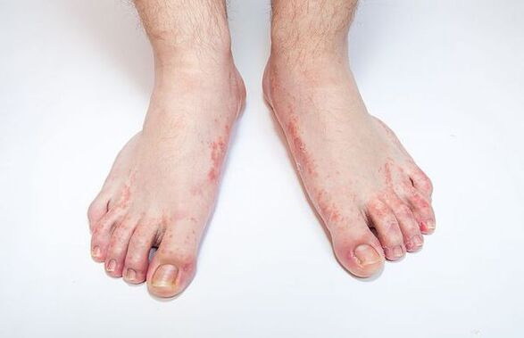
The first signs of foot mycosis appear in the form of itching and burning in the interdigital folds of the feet, the skin begins to peel, crack, turn red, and signs of swelling and inflammation appear.Complications can occur in the form of diaper rash and skin eczema.
Types of foot mycosis:
- erased - shown by moderate itching and hyperemia (redness) of the skin;
- acute - accompanied by severe itching and skin damage in the form of cracks;
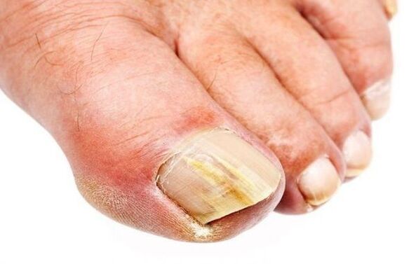
- nail mycosis (onychomycosis) - manifested by damage to the nail plate, which becomes thick and discolored;
- like a diaper - a crying area is formed;
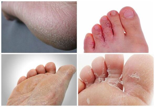
- squamous - lamellar scales appear;
- hyperkeratotic - accompanied by a rash in the form of papules and plaques on the arch of the foot;
- dyshidrotic - occurs with the development of swelling, weeping areas and blisters.
Pathogenesis of foot mycosis
The skin is the largest organ in the human body, accounting for 15% of the total body weight.It performs many functions, first of all, it protects the body from the effects of external factors of a physical, chemical and biological nature, from water loss, and also participates in thermoregulation.The skin consists of three layers: epidermis, dermis and subcutaneous fat.
The epidermis (outer layer of the skin) is the main barrier to fungi penetrating the skin.It is a multilayered squamous keratinized epithelium, which, in turn, consists of five layers and serves as a barrier.Keratinocytes are the main cells of the epidermis.It contains the protein keratin, which creates the outer layer of the skin and gives it elasticity and strength.Keratin cells in the epidermis are constantly exfoliated.
Dermatomycetes produce an enzyme - keratinase, which destroys keratin.Because of this, the fungus penetrates into the surface layer of the skin, where it continues to exist.The cell walls of dermatomycetes contain mannans, substances that can suppress local cellular immunity.The fungus T. rubrum, due to the action of manans, inhibits the proliferation of keratinocytes, as a result, desquamation of horny scales from the surface of the skin slows down and chronic infection develops.
Classification and stages of development of foot mycosis
Classification depends on the pathogen:
- Keratomycosis (pityriasis versicolor).
- Dermatophytosis (microsporia, superficial trichophytosis, foot mycosis, smooth skin mycosis, inguinal fold mycosis, onychomycosis).
- Candidiasis (candidiasis of the skin, nails).
- Deep mycoses (blastomycosis, sporotrichosis, chromomycosis).
Classification according to ICD-10:
- B35.1 - Nail mycosis.
- B35.2 - Mycosis of hands.
- B35.3 - Mycosis of the feet.
- B37.2 - Candidiasis of skin and nails.
Classification by localization:
- Cutaneous mycosis.
- Mycosis on the folds.
- Mycosis of the hands.
- Mycosis of the feet (squamous, hyperkeratotic, intertriginous, dyshidrotic forms).
- Onychomycosis (distal, superficial, proximal).
Classification by clinic:
- The form is deletedmanifests itself as peeling in the interdigital folds III–IV of the foot.Minor peeling may also occur on the soles and sides of the feet.
- Intertriginous formmanifested by hyperemia in the interdigital folds of the feet, and the appearance of foam is also possible, which causes the formation of erosion and cracks.Subjectively, itching and burning are observed.
- With a dyshidrotic formGroup blisters appear on the skin of the arch and lateral surface of the foot.More often they appear on healthy skin, then increase in size, merge and form larger multi-chamber blisters.When blisters open, erosions form.
- Squamous-hyperkeratotic formcharacterized by local or extensive thickening of the stratum corneum on the lateral and plantar surfaces of the feet.The affected skin area is covered with small scales like pityriasis.Exfoliation is especially noticeable in the skin folds.Cracks cause pain when walking.
Classification by clinic is very convenient from a practical point of view to determine further treatment tactics and monitor patients.
Based on the clinical picture of this disease, one can evaluate the causative agent of this disease.For example, the dyshidrotic form often occurs with foot mycosis caused by Trichophyton mentagrophytes var.interdigitale, the squamous-hyperkeratotic form is more often associated with T. rubrum, the chronic course and widespread processes are characteristic of opportunistic fungi Candida spp.and Aspergillus.
Complications of foot mycosis
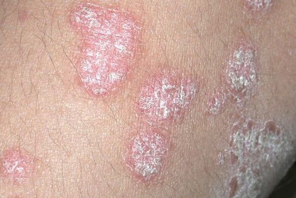
- Allergy to fungi.Under the influence of fungi, polyvalent sensitization is formed, that is, the body becomes more sensitive to fungal waste products, which are foreign to us and are strong allergens.The body reacts more sharply, which is shown by various rashes and skin reactions, chronic diseases of an allergic nature, such as skin eczema.The development or deterioration of pathologies such as bronchial asthma, allergic dermatitis, seborrheic dermatitis and psoriasis is possible.In addition, a person may more often experience complications of occupational allergies and drug intolerance.
- Pyoderma- pustular skin diseases (cellulitis, lymphangitis, phlegmon and osteomyelitis of the leg bones), which can lead to skin wounds that do not heal in the long term.Pyoderma occurs due to the fact that bacteria easily penetrate through erosion and cracks in the skin ("doors of infection").At the same time, the temperature rises, weakness and lethargy appear, which requires immediate surgical correction.
- Increased viral complicationsin the form of warts due to the presence of hyperkeratosis and cracks.The reason is a violation of the protective function of the skin, as a result of which it becomes more susceptible to any infection, including viruses.
- A general decrease in immunityand impaired microcirculation in the lower leg in patients with concomitant somatic diseases, such as diabetes mellitus and varicose veins.
- The spread of the disease to the nails and skin of the hands.When nail fungus occurs, they become deformed, ingrown nails, panaritium (purulent inflammation of the finger tissue), paronychia (inflammation of the periungual fold) and complete detachment of the nail plate are possible.
- Deterioration in quality of life.The acute form of foot mycosis is painful, making it difficult to wear shoes, and when lymphadenitis develops, they are accompanied by poor general health and fever.
Diagnosis of foot mycosis
The diagnosis of foot mycosis is based on the patient's complaints, medical history, clinical picture and laboratory results.Foot mycoses are among the diseases that necessarily require laboratory tests to confirm the clinical diagnosis.
The main method to confirm the diagnosis of foot mycosis is microscopic examination and culture.The material is a piece of skin, which is scraped from a lesion on the skin with a scalpel or glass;less commonly, the adhesive tape test is used.
Laboratory diagnosticsmycoses include microscopic examination and culture of material for fungi.Microscopic examinationis a real method for diagnosing pathogens, allowing one to identify the structure of the fungus within hours.Microscopic examination can reveal fungal elements in the form of mycelial threads and spores.The disadvantage of this method is that it is possible to obtain false positive and false negative results, which depend on many factors: the technique of taking the material, the peculiarities of storage and transportation, etc.
Cultural methodsis the most accurate diagnostic method, allowing one to identify the type of fungus to prescribe pathogenetic therapy.To prepare for the analysis, patients are not recommended to use any antifungal agents themselves for 1 month.
When prescribing systemic antifungal therapy, it is recommended tobiochemical blood testto determine the level of bilirubin, AST and ALT in relation to the need to monitor the function of the liver and bile ducts, as well as to prevent possible complications.
Differential diagnosis of foot mycosis:
- The squamous form is distinguished from psoriasis, eczema, and keratoderma.
- The interdigital form is distinguished from impetigo, diaper rash and candidiasis.
- The dyshidrotic form is distinguished from palmoplantar pustulosis.
Treatment of foot mycosis
Treatment should be carried out under the supervision of a dermatologist.
The main task in the fight against foot mycosis is timely detection, recognition and treatment before the development of nail fungus, which requires longer and more complex therapy (systemic antifungal therapy).At the same time, it is important to have effective drugs that match the modern clinical characteristics of foot mycoses.
Before starting treatment for a disease, dermatologists choose among possible treatment options.In most cases, medications are prescribed for topical use.The basis of treatment is the use of antifungal agents that have various effects.Medicines that stimulate blood circulation and medicines are also used to eliminate the main symptoms:
- Antifungal agent for external therapy: used externally 1-2 times a day for 4 weeks.
- In cases of significant hyperkeratosis of the feet, exfoliation therapy is first carried out: drugs from the group of azole derivatives, 1 time a day for 3-4 days, which act as keratolytics, that is, remove rough layers, thereby preparing the skin and increasing the penetration of antifungal agents into the dermis.
- If there is foam, use Castellani liquid;the solution is used externally 1-2 times a day for 2-3 days.Then the combined medicine is prescribed 2 times a day externally for 7-10 days.
- For severe itching, antihistamines are prescribed: histamine blockers H1-receptor – ethanolamine derivative 0.001 g 2 times a day orally for 10-15 days.
- Disinfect the shoes once a month until they are completely cured;you can use a spray whose active component is undecylenamidopropyltrimonium methosulfate.
- If the nail plate is affected, oral systemic antimycotic therapy must be prescribed for a period of 3 to 4 months.This therapy requires the supervision of a dermatologist, because the drugs themselves can lead to complications from internal organs, especially the liver, bile ducts, stomach, as well as the ineffectiveness of therapy and the formation of resistance to treatment.
It is necessary to treat foot mycosis, because if the fungus has settled on the skin, then without treatment it will not go anywhere, which means that the waste products of the fungus will always enter the surrounding tissue and blood, causing sensitization of the body and the development of chronic allergic diseases.
The presence of fungi indicates a decrease in immunity, and the skin damaged by mycosis practically does not perform a protective function.Therefore, all conditions are created for the addition of concomitant bacterial infection.
Patients with foot mycosis are an active source of infection for people around them and especially family members, so treatment in this case is an effective way to prevent fungal infections among healthy relatives and people around them.
A favorable environment for the development of fungal infections on the skin of the feet is a moist environment, so you should try to keep the skin of your feet dry.To do this, every evening you need to wash your feet with soap and dry your skin with a disposable paper towel, paying special attention to the space between your toes.
Forecast.Prevention
The prognosis for skin mycoses largely depends on the stage of the disease at which treatment is initiated.Therefore, if you notice changes in the skin, you should not delay your visit to the doctor.With timely and correct treatment of foot mycosispredictionfavorable: a complete recovery from the fungal infection occurs, the patient recovers.
If left untreated, the fungus can cause complications that not only disfigure the shape of the nails, but also affect the condition of the body as a whole.
Preventionfungal infection:
- Public prevention involves treating public places: baths, saunas, swimming pools, showers.Floors, appliances and household items must be disinfected.Staff and people who frequent public baths, saunas, etc.must undergo periodic preventive examinations.
Key personal precautions:
- observe personal hygiene rules when visiting public places;
- avoid damage and constant moisture on the skin and toenails;
- wear loose and comfortable shoes;
- Avoid contact with infected people.
Secondary personal prevention:
- maintain a hygiene regime for the skin of the feet;
- disinfect shoes, showers and bathrooms;
- increase immunity.